CNN-based Brain Tumor Classifier
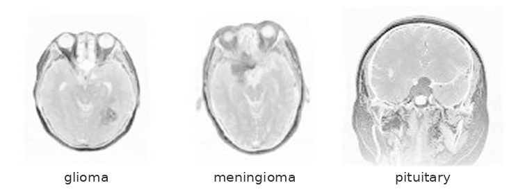
A convolutional neural network that classifies brain tumours from MRI into three types — glioma, meningioma and pituitary tumour.
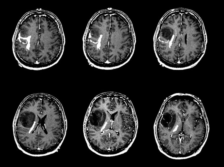
Conventional computer-aided diagnosis for brain tumours runs in three stages: find the tumour mass, segment it, then classify what you segmented. Every stage is a place to go wrong, and the segmentation step in particular has no universal solution across tumour location, shape and intensity.
This project tested whether the segmentation step is necessary at all.
Approach
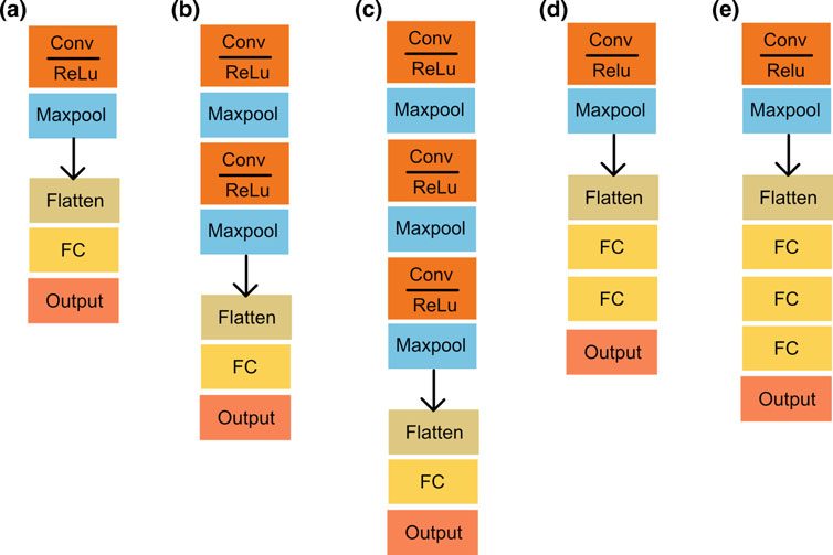
Five CNN architectures, all far simpler than AlexNet or VGG16, trained end to end on raw 64 × 64 images with no region-based preprocessing. Filter counts, pooling kernels and hidden-layer widths were held fixed so that only depth varied — the comparison isolates one variable.
Result
The winner was architecture 2 — two convolution/ReLU/maxpool blocks and one 64-neuron hidden layer — at 98.51% training and 84.19% validation accuracy.
It won for the right reason rather than the obvious one: it was the only architecture whose validation loss fell consistently across epochs. The others showed validation loss climbing while training accuracy improved, which is overfitting, and a useful reminder that on a few thousand medical images the headline accuracy figure means very little on its own. Widening the winner to 64 and 128 filters did not help either — capacity was not the binding constraint.
84.19% sits inside the 71.39–94.68% band that conventional region-based pipelines reach on the same dataset, without needing to find the tumour before classifying it.
Carried out at the Biomedical Research Group, Bandung Institute of Technology, and published at the World Congress on Medical Physics and Biomedical Engineering 2018.
Publications from this project
World Congress on Medical Physics and Biomedical Engineering, 2018
Brain Tumor Classification Using Convolutional Neural Network
Volume 1, pp. 183–189. Springer Singapore.
@inproceedings{abiwinanda2019brain,
  title     = {Brain tumor classification using convolutional neural network},
  author    = {Abiwinanda, Nyoman and Hanif, Muhammad and Hesaputra, S. Tafwida and Handayani, Astri and Mengko, Tati Rajab},
  booktitle = {World Congress on Medical Physics and Biomedical Engineering 2018},
  volume    = {1},
  pages     = {183--189},
  year      = {2019},
  publisher = {Springer Singapore}
}